Refractive Corneal Surgery: A Medical Advance of our Time
by Renato Alberto Meduri
Advanced techniques to reshape the curve of the cornea eliminating myopia, astigmatism and hypermetropia.
In the '70s, Fiodorov, a Soviet ophtalmologist, structurally
modified the curvature of the cornea to correct myopia, a medical advance
of our time.
To intuition, Americans have added technology by
substituting man's most stable hand and introducing constant and
dependable laser radiation. Modern refractive surgery allows the
reshaping of the curvature of the cornea, eliminating defects like myopia,
astigmatism and hypermetropia.
According to the defect, the cornea is
given a different anterior surface. It becomes more curved when correcting
hypermetropia, less curved or even concave when correcting myopia;
irregularities of astigmatism are eliminated homogenizing the rays of
curvature on the whole surface.
Correction is done by the laser
according to a program worked out by a computer on the basis of the
characteristics of the single eye, therefore according to a personalized
program.
The result is predictable with differences of less than half a
degree. It is highly improbable that the laser make a mistake. It has
three safety systems on watch at the same time, ready to stop the ray in
case of anomalous circumstances.
The procedure is now therefore
extremely reliable; the only real cause of failure could be an incorrect
evaluation on the part of the eye specialist about the information for the
intervention.
Not all eyes are adapted to be corrected either because
of improper structural characteristics or because of the presence of local
or systemic pathologies, which can interfere on the healing
process.
A visit to the eye specialist
It is therefore
mandatory to have an examination done by an eye specialist who will
examine in detail the characteristics of each eye: length, thickness,
surface and curvature of the cornea, refraction, and at the same time
general health condition as well as the family's medical history.
An
adequate examination requires to instill collyrium into the eyes, which
dilates pupils and blocks focus to see up close. No fear, everything comes
back to normal after a few minutes. The eye specialist takes down the
corneal surface's shapes and regularities by means of an instrument called
a topographer, which consists in producing on the cornea of the patient a
series of concentric light rings. A computerized optical system verifies
the regularity and curvature of the rings from which it calculates
curvature and regularity of the corneal surface.
Other ultrasound
instruments measure the thickness of the cornea in various points. This
exam is called pachimetry; moreover, ecobiometry is the evaluation of the
length of the eye.
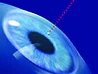
The excimer laser is then used. The computer
connected to the laser, and having received the previous measurements,
elaborates the corrective program. The program considers opening time and
the route the laser must travel on the surface of the cornea to reshape it
in a way to eliminate refractive errors. During the correction, the
patient must keep a steady fixation and if the laser finds that the eye
moves, it will automatically turn off.
The whole procedure lasts an
average of a few seconds and is absolutely pain free since the ocular
surface is desensitized with a few anaesthetic drops.
Immediately
after the operation, a therapeutic contact lense is placed and the patient
can leave the lab. Cortisone and antibiotic-based eye drops will be
prescribed and will be used for many days in order to allow problem free
healing of the treated corneal surface.
After the intervention, the
following must be avoided:
- exposure to irritating agents like dust,
smoke, wind;
- putting soap in the eyes during shower;
- swimming in
a pool or the sea for 15-20 days;
- rubbing the eyes vigorously;
protective lenses should always be worn.
There does not exit any
contraindications to watch TV or look at a monitor; the entertainment is
guaranteed.
If you have not thought about permanent make-up before
surgery, you will have to do without it for a few days until the cornea is
completely healed. For martial arts and boxing lovers, it is absolutely
forbidden to receive any blows or punches on the eyes during 15
days.
Before the surgery
It is important that the
eye be clean and uncongested and that the eyelids have no redness or
little crusts. It is advised to use antiseptic soap for the hands and face
during the days preceeding the surgery.
The mucous membrane, the
conjunctiva, is impaired and fragilized by difficult digestion, agitated
sleep and from exposure to irritating environments like smoke, dust and
ultraviolet rays. In such case, post-surgical recovery may not be optimal.
It is therefore advisable to follow a Franciscan or at least a healthy
lifestyle during the preceeding and immediately following days of the
intervention.
Various types of intervention
PRK: Reduction of myopia with excimer
laser.
The objective of PRK is to eliminate or reduce the use
of corrective lenses. It is achieved by modifying the cornea's
curvature, which is flat in the centre: in this way, light rays from
objects can focus clearly on the retina. The excimer laser vaporizes some
of the cornea's layers. The quantity of corneal tissue eliminated
determines how much myopia will be corrected.
Laser reduction of hypermetropia and
astigmatism.
Thermokeratoplasty with an holmium laser consists
in modifying the central curvature of the cornea, reshaping it. This is
achieved when the holmium laser ray hitting the cornea creates a
contraction and takes away the unwanted tissue.
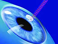
LASIK: surgical treatment of myopia coupled with
laser.
Also called Keratomileusis combined with excimer laser
treatment, LASIK is a mixed intervention.
In fact, after having made an
incision in the superficial layer of the cornea (epithelium) with a
microkeratome, the central part is flattened 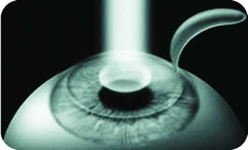 by the laser.
What is the laser?
The laser is a uniform and
powerful ray of light which can be produced in various ways. In
ophtalmology, various types of laser are used in order to take advantage
of the characteristics of each ray. The effect of laser radiation on the
target can be of evaporation, incision or heat.
What is the excimer laser?
The excimer laser
was developed at the end of the '70s and initially used to practice
extremely fine incisions on printed circuits.
For use in the field of
surgery, the produced light ray is controlled by a computer. It can remove
microscopic parts of the tissue on which it is directed with extreme
precision, since the ray's energy cuts the links between molecules and
creates an evaporation of the target without damage to the surrounding
tissues. The correction of refractive errors is done by bringing the
superficial layers in the central part of the cornea, modifying directly
the curvature and the optical power of the most important area of
focus.